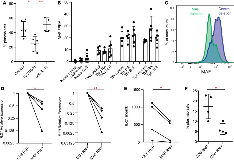
Figure 8. Tph cells induce B cell responses in an IL-21– and MAF-dependent manner.
(A) Quantification of plasmablasts among B cells in cocultures of memory B cells from controls cocultured with Tph cells from SLE patients with neutralization of either IL-21 or IL-10 (n = 3 donors for IL-21R-Fc, n = 2 donors for anti–IL-10, 2 replicates per donor). (B) Expression of MAF in RNA-seq data in indicated T cell populations. (C) Flow cytometric detection of loss of MAF expression in CRISPR/Cas9-treated CD4+ cells. (D) Expression of IL21 and IL10 by qPCR in CD4+ T cells after treatment with MAF- or CD8-targeting Cas9 complexes. Values plotted were normalized to CD8 targeting control. (E) Expression of IL-21 by ELISA in CD4+ T cells after treatment with MAF- or CD8-targeting Cas9 complexes. (F) Plasmablast generation in cocultures of Tph cells treated with MAF- or CD8-targeting complexes cocultured with allogeneic memory B cells. Data pooled from 2 experiments with 2 different donors. Error bars show mean ± SD (A, B, and F). *P < 0.05, **P < 0.01. Paired t test statistics of normalized data in D and E. Unpaired t test statistics shown in F.

