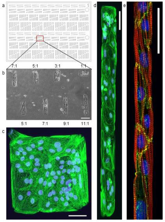
Fig. 2.
(a) Pattern consisting of rectangles with a range of aspect rations between 1:1 and 11:1. (b) A bright field image of 19–9-11 stem cell derived cardiomyocytes cultured on a portion of the pattern created with a Matrigel coated stamp using this pattern design. Aspect ratio is noted for each feature detailed above and below the image. Scale bar: 100 μm. (c, d) Fluorescent microscopy image of a 1:1 and a 9:1 feature. The nuclei are stained with DAPI (blue) and the protein α-actinin (green) highlights the sarcomere structure within the cells. (a-d) Scale bar: 50 μm. (e) Micropatterned lane with hESC-CMs cultured in Matrigel lanes on a 5kPa stiffness PDMS substrate for 18 days. Cell alignment and sarcomere organization shown in lanes of 20μm width. The nucleus is stained with DAPI (blue), the protein actin (red) highlights the internal organization of the cell, and the protein N-Cadherin (green) highlights the cell-cell interfaces. Scale bar 50 μm.

