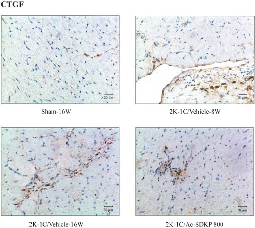
Figure 6.

Immunohistochemical staining for CTGF (with hematoxylin counterstaining) in LV of 2K-1C rats treated with Ac-SDKP. Brown color represents positive staining. CTGF-positive cells were observed in vascular endothelial cells and interstitial cells and were increased 8 and 16 weeks after clipping, whereas Ac-SDKP reversed this increase.
