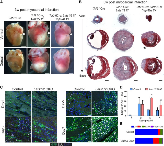
Figure 5.
Lats1/2 are required for cardiac scar maturation and compaction following myocardial infarction. (A) Gross heart morphology 3 wk after MI in control, Lats1/2 CKO, and Tcf21-iCre; Lats1/2 f/f; Yap/Taz f/+ mice. Suture used to ligate the left anterior descending coronary artery (LAD) shown with white arrow. Scale bar, 2000 µm. (B) Serial sections treated with Masson's trichrome stain 3 wk after MI in control, Lats1/2 CKO and Tcf21-iCre; Lats1/2 f/f; Yap/Taz f/+ mice. Red color tissue is muscle, and blue color is collagen (fibrotic tissue). Black arrows highlight myocardial muscle tissue. Scale bar, 1000 µm. (C) Representative images of 24 h EdU-labeling of control and Lats1/2 CKO mice after MI. Samples were pulse-chased with EdU (white) and cardiac fibroblasts were labeled with GFP (green). Nuclei stained with DAPI (blue). Scale bar, 25 µm. (D) Quantification of cardiac fibroblast proliferation dynamics after myocardial infarction. Representative image of experiment shown in Figure 5C. Statistical significance was determined by Mann–Whitney U test (P-value <0.1). (E) Stacked bar plot showing percentage of cells within each phase of the cell cycle as determined by flow cytometry analysis. See also Supplemental Figure S4B,C.

