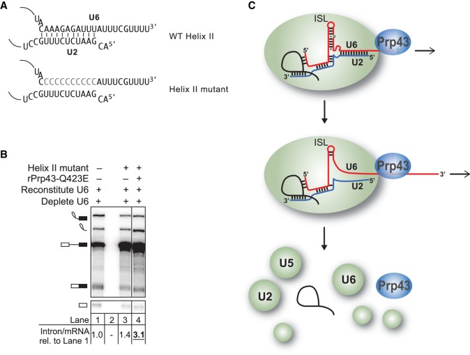
Figure 7.
A model for Prp43p function on the 3′ end of U6 involving disruption of structures beyond U2/U6 helix II. (A) U2/U6 helix II is depicted in the context of wild-type U6 or U6 with mutations that would disrupt base-pairing to U2. (B) Disruption of U2/U6 helix II does not bypass the requirement for Prp43p. In vitro splicing reactions were executed using radiolabeled ACT1 pre-mRNA in extracts (yJPS866) reconstituted with wild-type or helix II mutant U6 (illustrated in A) and with or without rPrp43p-Q423E, and then analyzed by denaturing PAGE. Quantitation of intron turnover is shown below the gel, wherein the ratio of excised intron to mRNA was calculated and normalized relative to the wild-type U6 control in lane 1; values with a substantial increase of twofold or greater over wild type are indicated in bold, underlined. (C) A mechanistic model for Prp43p function in splicing termination. See text for details.

