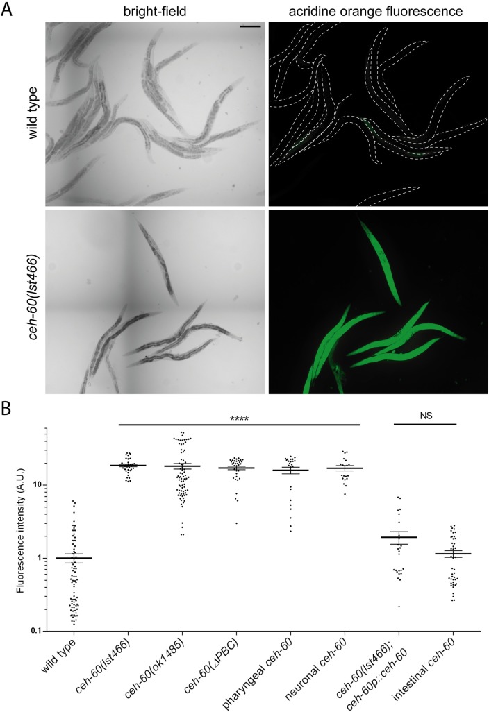
Fig 4. ceh-60 mutants have a more permeable cuticle.
(A) Representative images of acridine orange staining in wild-type and ceh-60(lst466) animals. Dotted white lines in wild-type acridine orange images show worm outline. Scale bar = 200 μm. (B) Acridine orange stains ceh-60 mutants but not wild-type animals. Expressing wild-type ceh-60 under the control of its own promoter (ceh-60p::ceh-60) or under an intestinal promoter (elt-2p::ceh-60) rescues the defect in ceh-60(lst466) mutants, but neuronal (odr-1p::ceh-60), pharyngeal (myo-2p::ceh-60), or PBC-truncated (ceh-60p::ceh-60(ΔPBC)) expression does not. Fluorescence intensity is shown on a logarithmic scale for clarity. ****p < 0.0001. N ≥ 20. Underlying data are available in S1 Data. A.U., arbitrary unit; NS, not significant.

