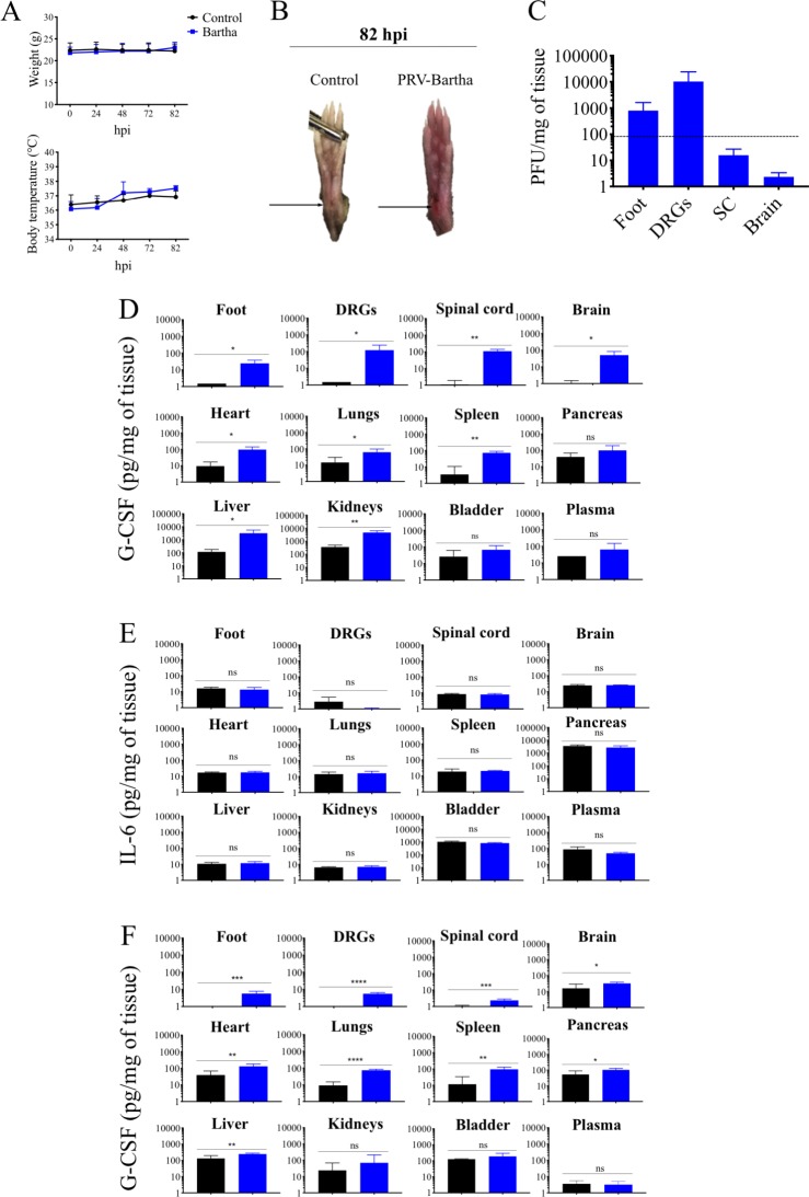
Fig 6. PRV-Bartha replicates efficiently in DRGs and partially induces a neuroinflammatory response in IFNAR KO mice.
(A) Body weight and temperature of IFNAR KO mice following PRV infection with Bartha strain (108 PFU) (blue) or control (black). (B) Representative images of mouse right hind paws at 82 hpi. Black arrows indicate the site of abrasion. (C) PRV DNA was quantitated in mouse tissues by qPCR using UL54 primers. PRV-Bartha loads are expressed as plaque forming units (PFU) per mg of tissue. (D) G-CSF and (E) IL-6 protein levels detected in PRV-Bartha infected and control mouse tissues at 82 hpi. (F) G-CSF protein levels detected in PRV-Bartha infected and control mouse tissues at 7 hpi. Protein levels were quantified by ELISA and expressed as pictogram (pg) per milligram (mg) of tissue. Two independent experiments were performed. Five mice per group were used per experiment.*, P< 0.05; ** P< 0.01; *** P< 0.001; ****P< 0.0001; ns, not significant.

