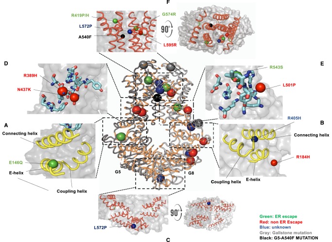
Figure 1. Structural motifs and missense variants of ABCG5/G8.
The crystal structure of ABCG5/G8 (PDB accession number: 5DO7) is plotted in cartoon presentation, with conserved amino acids colored in light orange (center). Residues whose missense mutations cause Sitosterolemia are highlighted in colors based on their maturation in cells. Green: ER-escape; red: non-ER-escape; blue: unknown. Residues whose missense mutations cause gallstone and other lipid phenotypes are plotted in gray. Only three can be localized in the current crystal structure. A540 on ABCG5 is highlighted in black. G5: ABCG5; G8: ABCG8. (A and B) The triple-helical bundle. (C) The NBDs of both subunits. (D and E) Stick presentation of the polar amino acids in the polar relay motif. (F) The ECD and the putative sterol-binding/entry site where the α-carbon of G5-A540 is highlighted in black. All figures are generated by using PyMOLTM.

