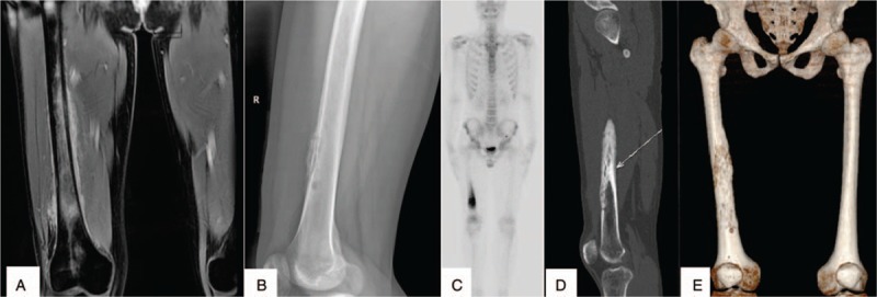
Figure 1.

X-radiography, high-resolution computed tomography, and 3-dimensional reconstruction of the bone showing multiple lucent defects with a moth-eaten destruction of osteolysis in the right femur (A–C). Emission CT showed significantly increased uptake in the femur (D). Magnetic resonance imaging shows abnormal signal shadow in the femoral bone marrow cavity (E).
