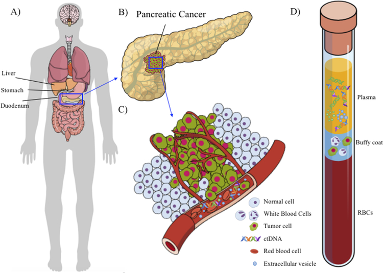
Figure 1. Pancreatic cancer and liquid biopsy (blood).
A) A schematic of the human body and abdominal cavity that shows pancreas is hidden in the abdominal cavity behind stomach, under the liver. B) A schematic of a clump of pancreatic cancer in the pancreas. This is a representative schematic, and the clump could be in any location in the pancreas. C) A schematic of the dissemination of pancreatic cancer components (ctDNA, exosomes, and CTCs) into the blood stream. D) A schematic of a blood tube with three main compartments—plasma, buffy coat, and red blood cells—each containing pancreatic cancer components.

