
Figure 2.
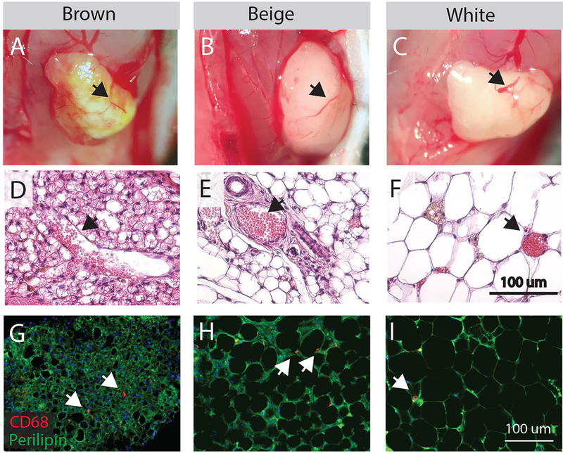
Characterization of transplanted fat engraftment. (A-C) Gross images of fat 14 days post-transplant on the surface of the supraspinatus muscle following removal of the overlying trapezius muscle. Brown, beige and white grafts have visible red vessels that bleed when cut (arrows). (D-F) Vessels identified on H&E stained sections of fat grafts (arrows) contain red blood cells (pink circles). Adipocytes in brown and beige fat exhibit multilocular morphology (multiple lipid droplets per cell). (G-I) CD68 staining for macrophages in fat grafts reveals a diffuse distribution of positive cells (arrows) amongst perilipin positive adipocytes.