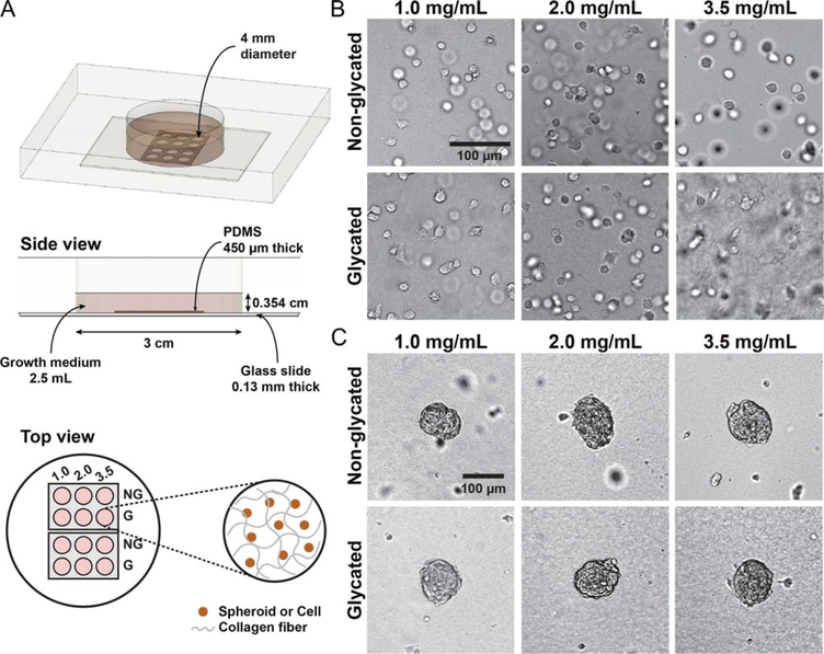
Figure 1.
An array microwell device for studies of 3D tumor cell invasion. (A) Experimental setup. A Plexiglas plate with a cylindrical hole of 3 cm in diameter and 1 cm in depth is sealed at the bottom with a No. 1 glass slide using high vacuum grease. Two PDMS array devices are plasma-bonded to the surface of the glass slide. Each array device contains a 450 μm thick PDMS membrane with 6 wells that are 4 mm in diameter. Cell-embedded collagen is placed in each of the wells. Typically, the collagen thickness is about 400 μm and the medium above the collagen is about 3.14 mm in height. (B) Bright-field images of MDA-MB-231 cells embedded in glycated and non-glycated collagen gels. (C) Bright-field images of MDA-MB-231:MCF-10A (1:1 ratio) spheroids embedded in glycated and non-glycated collagen gels. In each micro-well array device, six different collagen types are used: glycated and non-glycated collagen at concentrations of 1.0, 2.0, and 3.5 mg/mL. These images are taken at t = 0, which is approximately 2 h after the cells or spheroids have been introduced into the collagen.

