Figure 2.

OTC protects neuronal cultures against OGD-caused cell injury
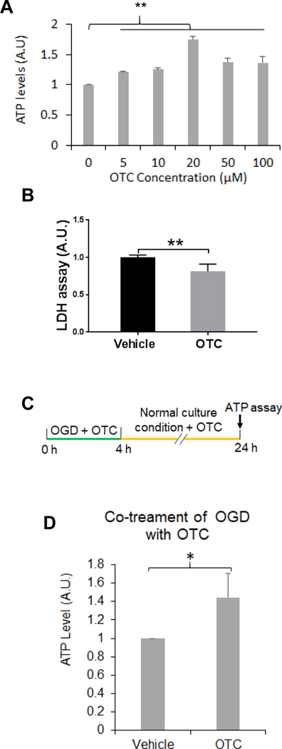
A. OTC increases striatal cell viability following OGD treatment. The striatal cells were incubated with a buffer free of glucose containing the indicated concentrations of OTC in a chamber free of oxygen. After l4 h, the cells were collected for measuring ATP levels. Data are shown as mean ± SD. N = 3; ** p < 0.0l, F(5, 12) = 66.67, one-way ANOVA with Tukey’s multiple-comparison test: p 0 μM vs 5 μM = 0.003, p 0 μM vs l0 μM = 0.0007, p 0 μM vs 20 μM < 0.000l.
B. OTC decreases OGD-induced LDH release. Striatal cells were treated with OGD in presence of 20 μM of OTC or vehicle and after 14 h, LDH release assay was performed. Data are shown as mean ± SD, n = 3; ** p < 0.01 (two-tailed paired Student’s t-test).
C. Diagram of the experimental design for co-treatment of OGD with OTC as shown in (D). Primary mouse cortical neurons were treated with OGD along with 20 μM OTC and after 4 h, the cells were cultured in the normal condition and continued to receive 20 μM OTC treatment until 24 h before ATP was measured.
D. Co-treatment of OGD with OTC increases neuronal viability in primary mouse cortical neuronal culture. Data are shown as mean ± SD, n = 3; p = 0.0413, t(4) = 2.966, two-tailed unpaired Student’s t-test.
