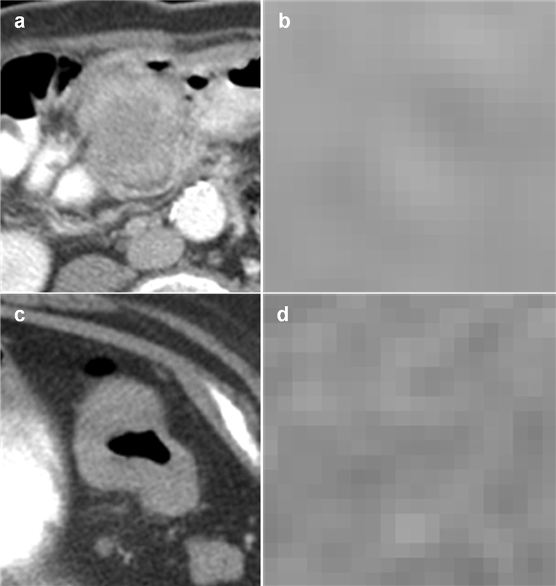
Fig. 3.
Examples of MSI positive and negative tumors classified correctly by our prediction model. (a) shows the cross-sectional axial image of an MSI tumor and (b) shows the tumor at the pixel level. (c) shows the cross-sectional axial image of an MSS tumor and (d) shows the tumor at the pixel level. Homogeneity is measured in MSI tumors which tend to be more indolent than MSS tumors. This is consistent with radiomics of other cancers indicating tumor heterogeneity is a marker for disease aggressiveness

