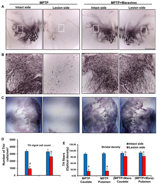
Figure 7. Oral maraviroc treatment protects dopaminergic neurons in the SNpc and THir fibers in the striatum of hemiparkinsonian monkeys.
Naïve female rhesus monkeys received a right intracarotid injection of MPTP. After 7 d of injection, monkeys displaying classical parkinsonian postures received maraviroc (1 mg/kg body wt/d) orally mixed with banana. MPTP group of monkeys also received normal banana as control. A) After 30 d of treatment, nigral sections were immunostained for TH. B) A magnified image of TH-stained SNpc is shown. C) Striatal sections were also immunostained for TH. D) Estimates of dopaminergic nigral cell number [MPTP, n=4; (MPTP+maraviroc), n=4] were performed bilaterally using an unbiased design-based counting method (optical fractionator, StereoInvestigator; Microbrightfield, Williston, VT). All counting were performed by a single investigator blinded to the experimental conditions. ap < 0.001 vs MPTP-intact; bp < 0.01 vs MPTP-lesion. E) Optical density of TH fibers in caudate and putamen was calculated by digital image analysis. Six different striatal sections of each monkey were analyzed. ap < 0.001 vs MPTP-intact; bp < 0.01 vs MPTP-Caudate-lesion; cp < 0.01 vs MPTP-Putamen-lesion.

