Figure 3.

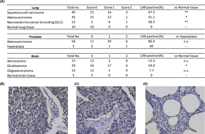
Coxsackievirus and adenovirus receptor (CAR) expression in various human tumor tissues. A, CAR expression in human lung, prostate, and brain tumors. Immunohistochemical staining was carried out on cancer tissue arrays using CAR antibody H‐300. Estimated visual intensity of CAR immunostaining was graded on an arbitrary three‐point scale: negative (0), positive (1), strongly positive (2). CAR, coxsackievirus and adenovirus receptor; SCLC, small cell lung cancer. Statistical analysis was done by Mann‐Whitney U test (*P < .05, **P < .01, n.s., not significant). B‐D, Representative CAR staining in the lung cancer tissue array. B, SCLC (score 2). C, SCLC (score 1). D, Normal lung tissue (score 0). All data were documented under a magnification of ×400
