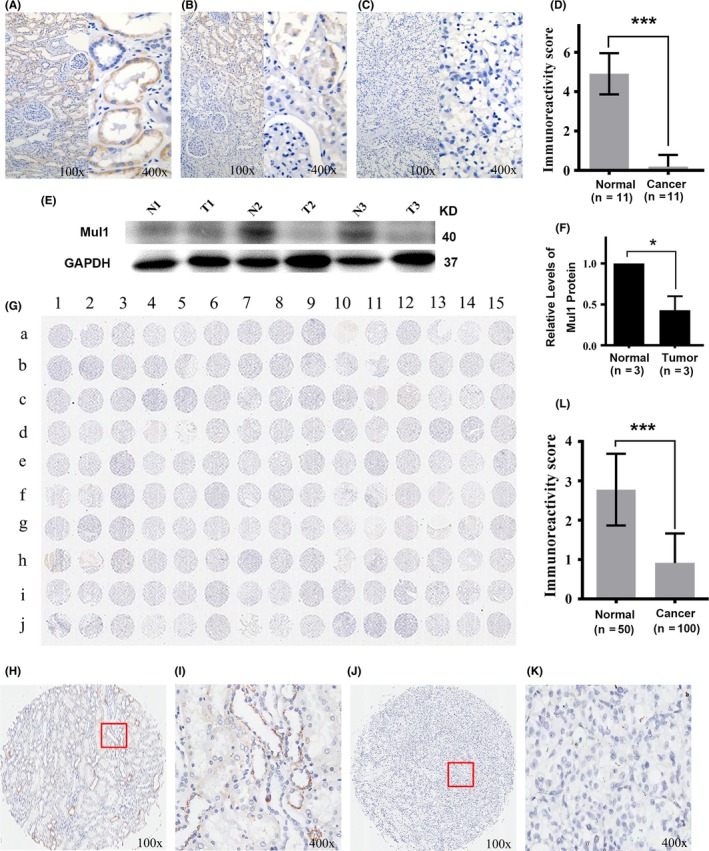
Figure 1.

Expression of mitochondrial E3 ubiquitin ligase 1 (Mul1) protein in clear cell renal cell carcinoma (ccRCC) tissues and their adjacent normal tissues. (A‐C) Representative images showing adjacent normal tissues expressing high levels (A), tissues near tumor tissues expressing intermediate levels (B) and ccRCC tissues expressing low levels of Mul1 (C) collected from 11 patients with ccRCC in our hospital. Left half is an image with low‐resolution (100×) and right half with high‐resolution (400×). (D) Plot showing a comparison of immunoreactivity scores of Mul1 between normal and cancer tissues from ccRCC patients as stated above. Mean and SD for normal tissues are 4.91 ± 1.04 and those for cancer tissues are 0.18 ± 0.60. ***P ≤ .001. Sample size is 11. (E,F) Immunoblot assays (E) and quantification (F) showing levels of Mul1 in tumor (T1, T2 and T3) and their respective adjacent normal tissues (N1, N2 and N3) from three ccRCC patients from our hospital. *P ≤ .05. (G) Full immunohistochemical image showing expression levels of Mul1 in a tissue microarray containing 100 ccRCC and 50 non‐cancerous tissues. (H‐K) Representative images showing adjacent normal tissues (H,I) and ccRCC tissues (J,K) with low‐resolution (100×) (H,J) or high resolution (400×) (I,K). (L) Plot showing a comparison of immunoreactivity scores of Mul1 between normal (n = 50) and cancer tissues (n = 100) as shown in G. Mean and SD for normal tissues are 2.78 ± 0.91 and those for cancer tissues are 0.91 ± 0.75. ***P ≤ .001
