Figure 2.

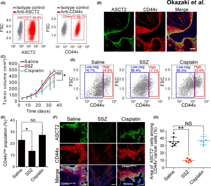
ASCT2+/CD44vhigh head and neck squamous cell carcinoma (HNSCC) cells are highly sensitive to xCT inhibition. A, Flow cytometric analysis of ASCT2 and CD44v expression in HSC‐2 cells. FSC, forward scatter. B, Immunohistofluorescence staining of ASCT2 (green) and CD44v (red) in tumors formed in athymic mice injected with HSC‐2 cells. Nuclei in the merged image were stained with 4ʹ,6‐diamidino‐2‐phenylindole (DAPI, blue). Scale bar, 50 μm. C, Volume of tumors formed by HSC‐2 cells in nude mice treated with saline (control), sulfasalazine (350 mg/kg per day) or cisplatin (2 mg/kg every 3 days). Data are means ± SD for 5 mice per group. *P < 0.05 (Student's t test); NS, not significant. D, Flow cytometric analysis of CD44v expression in lineage marker‐negative cells isolated from tumors formed by HSC‐2 cells in nude mice treated with saline, sulfasalazine or cisplatin as in (C). E, Quantification of CD44vhigh tumor cells as in (D). Data are means ± SD for 3 mice per group. *P < 0.05 (Student's t test). F, Immunohistofluorescence staining for ASCT2 (green) and CD44v (red) in tumors formed by HSC‐2 cells in nude mice treated as in (C). Nuclei in the merged images were stained with DAPI (blue). Scale bars, 50 μm. G, Quantification of the area occupied by ASCT2+ cells in the CD44v‐expressing region of tumors formed by HSC‐2 cells in nude mice as in (F). Each data point corresponds to an individual field of view, and bars indicate mean ± SD values. **P < 0.01 (Student's t test)
