Figure 3.

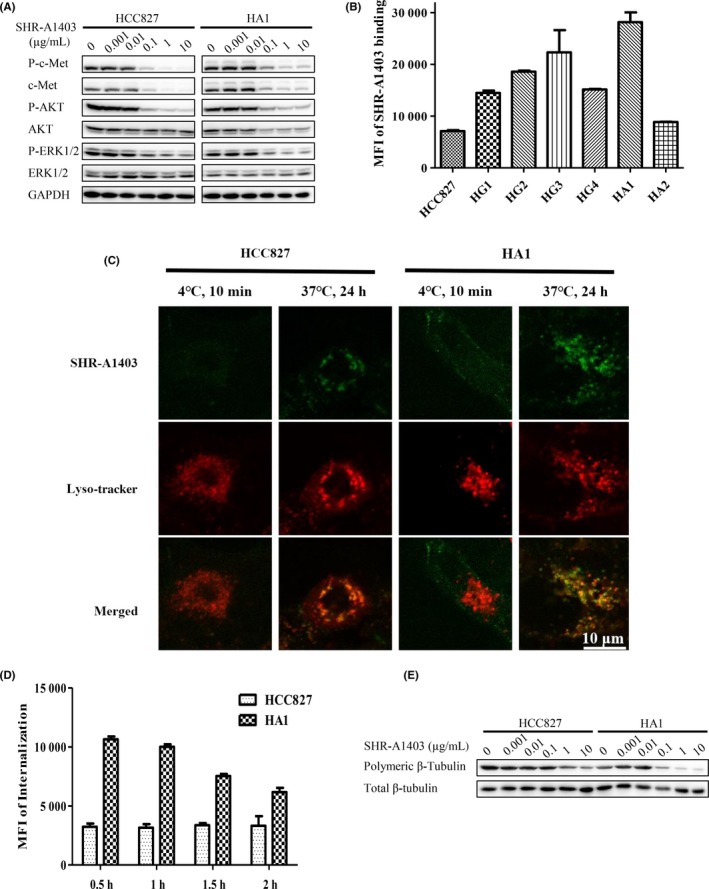
Antitumor activity of SHR‐A1403 is dependent on c‐mesenchymal‐epithelial transition factor (c‐Met) expression levels. A, HCC827 and HA1 cells were treated with different concentrations of SHR‐A1403 for 24 h, and whole‐cell lysates were analyzed by western blotting. B, Binding affinity of DyLight 488 NHS‐ester‐labeled SHR‐A1403 to parental and resistant HCC827 cells was measured by flow cytometry. Representative results are shown. C, Colocalization of SHR‐A1403 (green) with lysosomes (red). Parental and resistant HCC827 cells were incubated with DyLight 488 NHS‐ester‐labeled SHR‐A1403 (1 μg/mL), and lysosomes were labeled with Lyso‐Tracker Red. Samples were analyzed under confocal microscopy. D, Internalization of SHR‐A1403 in HCC827 and HA1 cells. Cells were incubated with DyLight 488 NHS‐ester‐labeled SHR‐A1403 (1 μg/mL) as described in Materials and Methods, and internalized fluorescence was analyzed immediately by flow cytometry. Representative images are shown. E, Effects of SHR‐A1403 on tubulin polymerization in AZD9291‐resistant HCC827 and HA1 cells. Cells were incubated with different concentrations of SHR‐A1403 for 48 h, then intracellular polymeric β‐tubulin and total β‐tubulin were analyzed by western blotting
