Fig. 7.
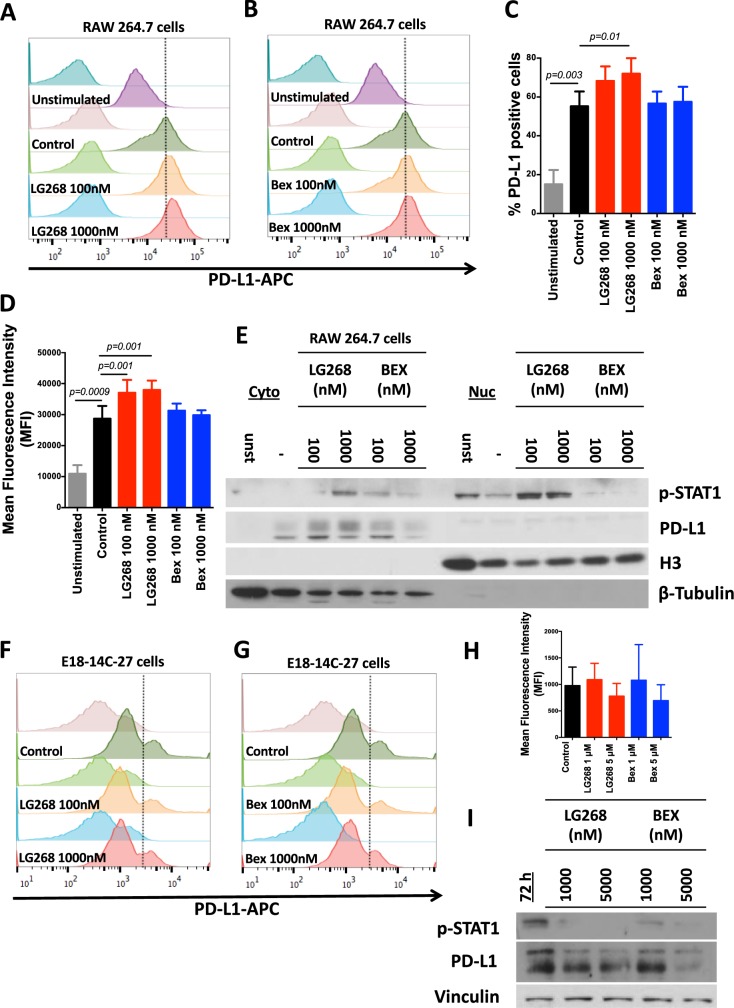
LG268 treatment increases the expression levels of PD-L1 in macrophages. RAW 264.7 macrophage-like cells were treated with conditioned media from E18-14C-27 breast cancer cells isolated from a mammary tumor in a MMTV-Neu mouse; representative histograms for levels of PD-L1 after treatment with LG268 a or bexarotene b for 24 h, are shown. Quantification of the percentage of positive PD-L1 (F = 7.74; df = 5) c or mean florescence intensity (F = 9.765; df = 5) d for four independent experiments. e Cytoplasmic (Cyto) vs. nuclear (Nuc) expression of PD-L1 and p-STAT1 in RAW 264.7 macrophage-like cells after stimulation with conditioned media from E18-14C-27 cancer cells. Representative histograms of PD-L1 expression in E18-14C-27 cancer cells after treatment with LG268 f or bexarotene g for 72 h. h Mean florescence intensity of E18-14C-27 cancer cells positive for PD-L1. i E18-14C-27 cancer cells were treated with LG268 or bexarotene for 72 h and levels of PD-L1 and p-STAT1 were analyzed by western blot. n = 3–4, Bars represent average and error bars the standard error of the mean (SEM)

