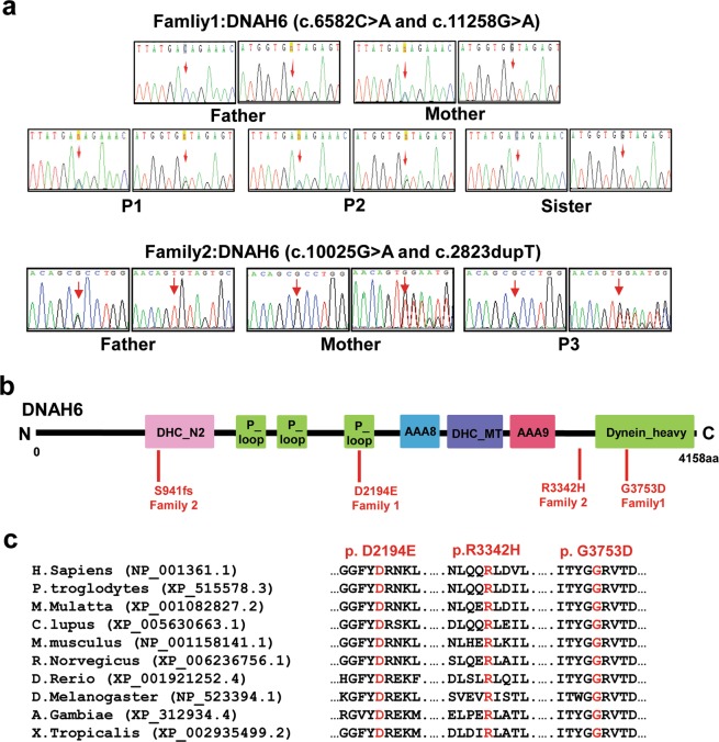
Figure 2.
Segregation analysis and functional characterization of the DNAH6 mutation. (a) Sequence chromatograms for DNAH6 (c.6582 C > A and c.11258 G > A) in family 1, and DNAH6 (c.10025 G > A and c.2823dupT) in family 2. Both unaffected parents were heterozygous, whereas the patients (P1, P2, and P3) were compound heterozygous for these mutations and the unaffected sister harbored the wild-type variant. (b) Structure of DNAH6 protein; predicted functional domains are shown together with the position of novel mutations identified in MMAF patients. (c) Cross-species alignment showing strong conservation at positions 2194 and 3753 in DNAH6.

