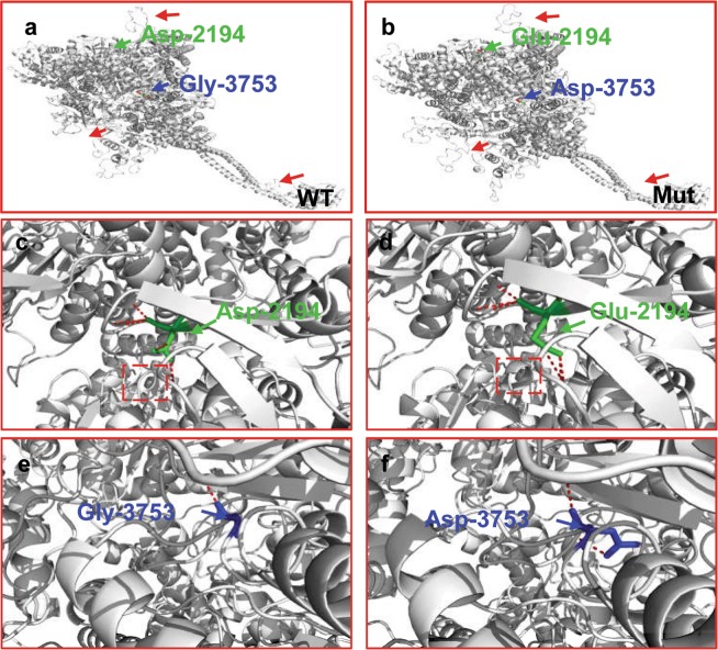
Figure 3.
Modeling of wild-type and mutant DNAH6. Molecular structures of wild-type and mutant DNAH6 proteins were modeled with SWISS-MODEL software based on the template of dynein heavy chain, cytoplasmic (3vkh.pdb). (a,b) Structures of wild-type and mutant DNAH6 proteins; β-sheets (red arrows) are altered compared to the wild-type protein. (c,d) Enlarged view of the Asp-2194 site in wild-type and mutant DNAH6 protein. (e,f) Enlarged view of the Gly-3753 site in wild-type and mutant DNAH6 protein. Both Asp2194Glu and Gly3753Asp mutations of DNAH6 altered the structure of the protein. The positions of mutations are highlighted in blue.

