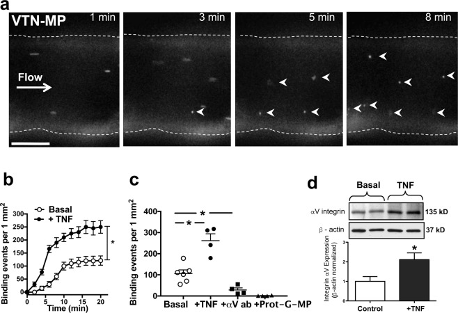
Figure 1.
Microparticle (MP) binding under flow in the isolated middle cerebral artery. (a) Fluorescence images of representative time-lapse recording demonstrate firm binding of VTN-MPs (0.1 µg/ml) on the luminal surface of the isolated mouse MCA in the presence of physiological pressure and flow (white arrowheads show stationary MPs). Scale bars: 50 μm. (b) Summary data (n = 4–6) show the rate and number of binding events of VTN-MPs in the absence (basal) or presence of TNF (7 ng/ml, 4-hour), or after incubating with αV integrin blocking antibody, or after delivery of Protein G conjugated MPs (c,d) Representative images and summary data of Western immunoblot shows increased expression of αV integrin on TNF-exposed mouse MCA. Data are shown as mean ± S.E.M; *p < 0.05.

