Figure 2.

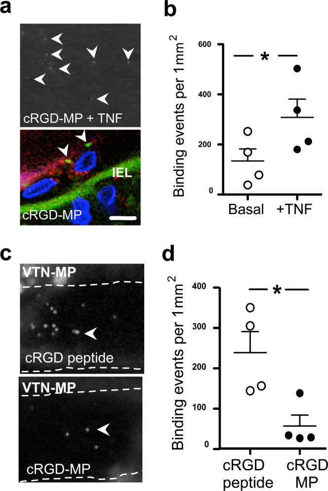
Modulation of RGD-endothelial interactions by microparticle-carried ligands. (a) Representative images show localization of cRGD-MPs (white arrowheads - upper panel; shown in green with arrowheads - lower panel) and CD-31 staining (red - lower panel) on TNF-activated endothelial cells (blue - DAPI). (b) Bindings of cRGD-MPs (0.1 µg/ml) in isolated, pressurized MCA, before and after incubation with TNF. (c) Representative images show VTN-MP bindings in TNF-exposed MCA with prior intraluminal delivery of cRGD peptide (100 μg/ml) or cRGD-MPs (0.1 µg/ml). (d) Summary data of VTN-MP bindings in TNF-exposed MCA with cRGD peptide or cRGD-MPs. IEL: Internal Elastic Lamina. Scale bar: 10 μm. Data are mean ± SEM; *p < 0.05.
