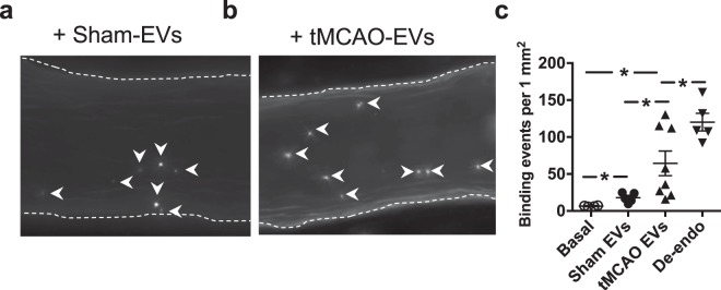
Figure 5.
RGD protein bearing extracellular vesicles (EVs) promote platelet adhesion under flow. (a,b) Fluorescence images of representative time-lapse recording demonstrate firm binding of CFSE-labelled platelets on the luminal surface of the isolated mouse MCA pre-treated with EVs isolated from sham and tMCAO animals, and in the presence of physiological pressure and flow (white arrowheads show stationary platelets). (c) Summary data show adhered platelets (white arrow heads) under basal condition, and after the endothelium was removed with air (De-endo), or when sham EVs or EVs from tMCAO were delivered prior to platelets. Data are mean ± SEM, *p < 0.05.

