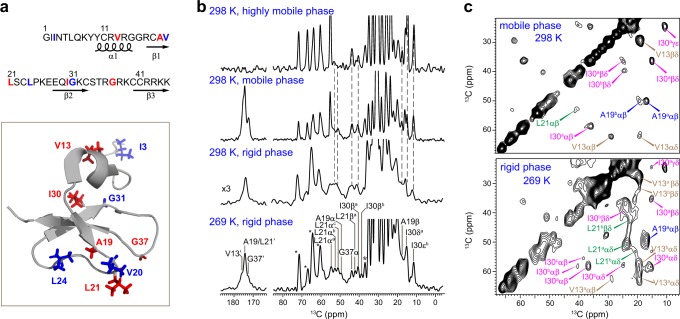
Fig. 1.
Isotope-labeling schemes and 13C solid-state NMR spectra of hBD-3 analog. a The 13C, 15N-labeled residues are in red for VALIG peptide, and in blue for IVLG peptide in both the amino acid sequence and solution monomeric structure of wt-hBD-3 (PDB 1KJ6). b 1D 13C spectra of VALIG peptide in POPC/POPG bilayers at 269 K and 298 K. From top to the bottom are INEPT, DP, and CP spectra measured at 298 K, and CP spectrum at 269 K. Dashlines indicate the resolved peptide peaks. c 2D 13C-13C correlation spectra of VALIG with 100-ms DARR mixing. The rigid and mobile components are selected using 1H–13C CP and 13C DP, respectively. Greek letters indicate carbon sites and superscripts annotate conformers. I30aαβ: carbon α to carbon β cross peak of subtype-a I30

