Fig. 5.
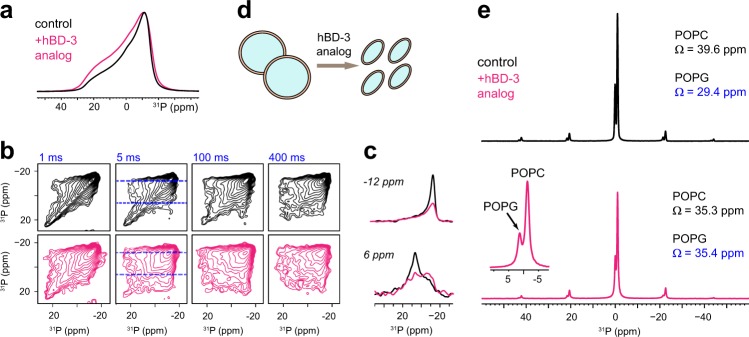
The hBD-3 analog perturbs membrane morphology and rigidifies negatively charged lipids. a 31P static spectra of control POPC/G membranes without (black) and with (magenta) hBD-3 analog at 298 K. b 2D static 31P–31P exchange spectra measured with 1, 5, 100, and 400 ms mixing times. The hBD-bound sample (magenta) has more off-diagonal signals due to rapid exchange. c Cross-sections at 6 ppm and 12 ppm, with diagonal normalized (asterisk). d Illustration of the effect of hBD-3 analog on POPC/G vesicles. e 1D MAS 31P spectra shows resolved POPC and POPG signals. Fitting the sideband patterns provides information on the chemical shift anisotropy. Peptide-bound POPG has an increased span (Ω) due to reduced motions of headgroups

