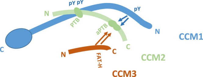
Figure 8.

Schematic representation of binding interaction among CCM proteins within CSC complex. Our current data suggests that CCM1 utilizes 2nd and 3rd NPXY motifs (pY) in its center portion to bind to CCM2 classic PTB domain (PTB). The remaining 1st NPXY motif competes with the CCM3 (FAT-H) to bind to the newly defined atypical PTB domain (aPTB) present at the C – terminus of the CCM2, suggesting CCM2 plays a central role in the CSC. CCM1: blue color; CCM2: light green color; CCM3: red color.
