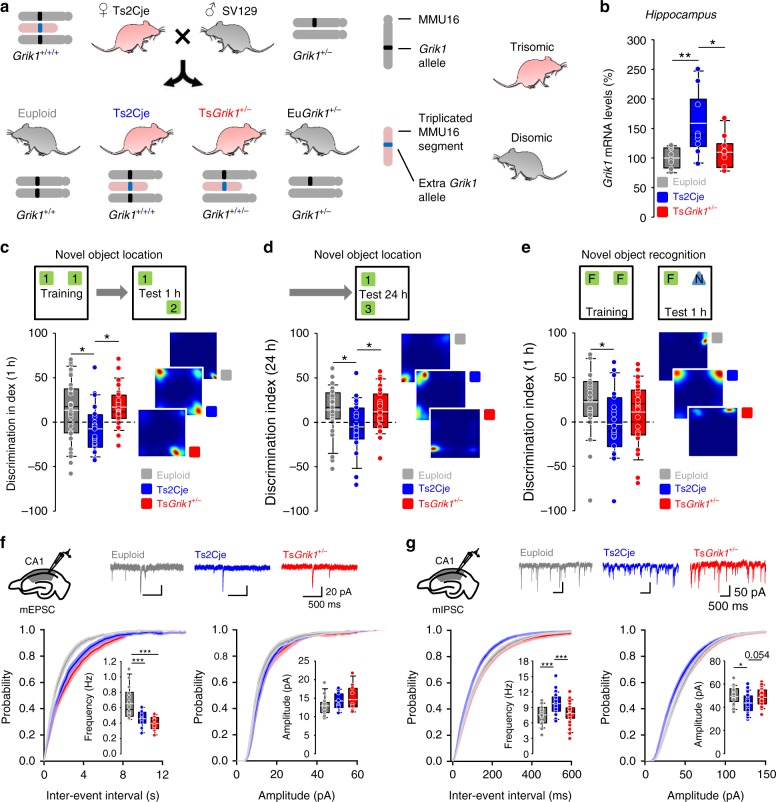
Fig. 1.
Grik1-dependent spatial memory impairment and anomalous basal inhibition in CA1 of Ts2Cje mice. a Experimental approach for Grik1 dose normalization. We crossed trisomic Ts2Cje females with Grik1+/− males, obtaining offsprings consisting of wild-type mice (Grik1+/+, euploid), trisomic mice with all genes in the extra segment triplicated (Ts2Cje), trisomic mice with all genes triplicated except Grik1 (TsGrik1+/−), and disomic mice monozygous for Grik1 (Grik1+/−). b Real-time qPCR quantification of hippocampal Grik1 mRNA levels in euploid, Ts2Cje, and TsGrik1+/− mice. Expression levels were normalized to those of euploid animals (n = 8 for euploid, n = 10 for Ts2Cje, n = 10 for TsGrik1+/−, one-way ANOVA+Holm–Sidak). c–e Performance in the novel object location (NOL) and novel object recognition (NOR) paradigms. A schematic representation of the paradigms appears at the top, whereas the bottom displays box plots showing the quantification of data for each test session, and heat plots tracking representative animals’ behavior. Ts2Cje mice did not discriminate between familiar and novel locations at short-term (1 h after training, c) and long-term (24 h after training, d) intervals in the NOL task. Normalization of Grik1 dosage in TsGrik1+/− mice restored the memory performance to euploid levels (n = 31 for euploid, n = 23 for Ts2Cje, n = 27 for TsGrik1+/−, one-way ANOVA+Holm–Sidak). e Ts2Cje mice presented impaired discrimination between familiar and novel objects in the NOR test 1 h after training. Grik1 dosage normalization partially rescued this impairment (n = 29 for euploid, n = 23 for Ts2Cje, n = 27 for TsGrik1+/−, one-way ANOVA+Holm–Sidak). f Miniature excitatory postsynaptic currents (mEPSCs) were recorded at −75 mV in hippocampal CA1 pyramidal neurons to evaluate basal excitatory synaptic transmission. The schematic representation of the electrode arrangement in the hippocampus and representative traces of mEPSC recordings are shown in the top. Bottom, cumulative distribution and box plots showing reduced mEPSC frequency (left) and unaltered amplitude (right) in Ts2Cje and TsGrik1+/− mice (n = 14 for euploid, n = 11 for Ts2Cje, n = for TsGrik1+/−, one-way ANOVA+Holm–Sidak). g Miniature inhibitory postsynaptic currents (mIPSCs) were recorded at −60 mV in CA1 pyramidal neurons to evaluate basal inhibitory transmission. Top, schematic representation of electrode arrangement and representative traces of mIPSCs. Bottom, cumulative probability and box plots showing increased frequency (left) and reduced amplitude (right) of mIPSCs in Ts2Cje mice. Normalization of Grik1 dosage recovered these phenotypes to euploid levels (n = 22 for euploid, n = 31 for Ts2Cje, n = 23 for TsGrik1+/−, one-way ANOVA+Holm–Sidak). *p < 0.05, **p < 0.01, ***p < 0.005. For detailed data values and statistics, see Supplementary Table 2

