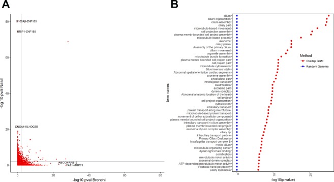
Figure 6.
Network analyses for the differentially expressed genes (DEGs). (A) Scatter plot of the GGM network edges (partial correlation) for the DE genes. Each red dot represents of an edge in bronchial tissue (horizontal axis) versus nasal tissue (vertical axis). In light black: the critical value at BH The figure displays the respective gene pairs for the most similar edges. (B) The 50 most significant GOs for the set of Overlapped genes. The panel displays the corresponding mean (p-values), and error bars represent +2 standard errors over the 500 random samples.

