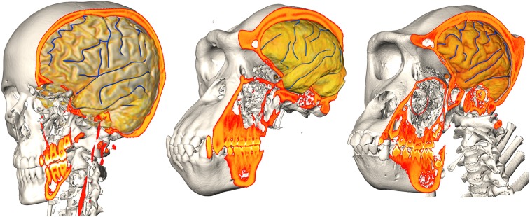
Fig. 1.
Same-subject coregistered CT/MRI datasets of a human (Left), chimpanzee (Center), and gorilla (Right). Surface reconstructions of bony structures were derived from CT data, while volume renderings of brain segmentations were obtained from postprocessed MRI data. Delineations of some of the brain sulcal features used in the study are shown in blue.

