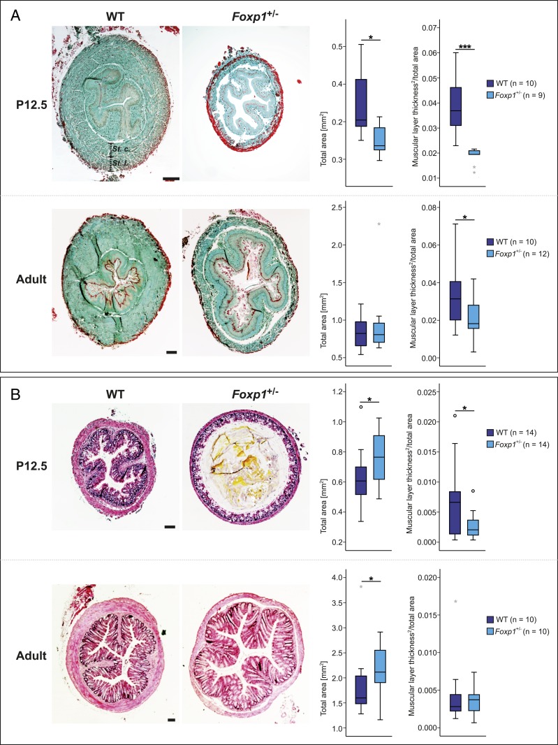
Fig. 2.
Tunica muscularis of the esophagus and the colon is altered in Foxp1+/− mice. (A) The esophagus is significantly smaller in Foxp1+/− animals compared with WT littermates. Sections from Foxp1+/− organs harvested at P12.5 and adult stage are 25 and 11% smaller compared with WT sections, respectively. The thickness of the tunica muscularis is significantly reduced in Foxp1+/− mice at both stages, by 52% at P12.5 and 40% at adult stage. (B) The colon of Foxp1+/− animals reveals a strong reduction in muscle layer thickness by 61% together with a dilated lumen at P12.5. In the adult colon, the total size of Foxp1+/− sections is significantly increased by 14% compared with WT sections, whereas morphological differences regarding the thickness of the muscular layer were not observed. A comparable number of male and female animals of both genotypes were used in the experiments. Esophageal sections were stained with Masson–Goldner trichrome, and colon sections were stained with hematoxylin and eosin. Asterisks indicate significant difference (*P ≤ 0.05, ***P ≤ 0.001; ANCOVA). (Scale bars, 100 µm.)

