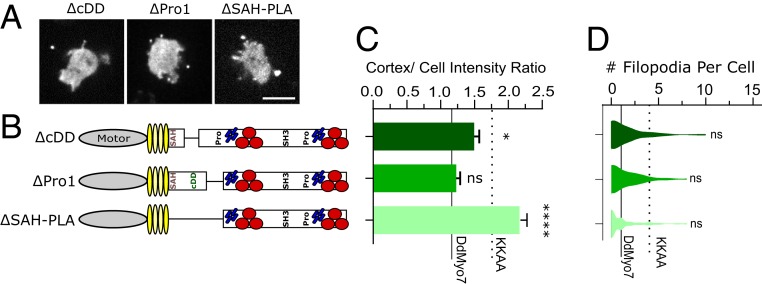
Fig. 2.
The proximal tail regulates DdMyo7 activity. (A) Micrographs of D. discoideum cells expressing GFP-tagged deletion constructs in myo7 null cells. (Scale bar: 10 μm.) (B) Schematic illustration of constructs. (C) Cortical band intensity of proximal tail deletion mutants. (D) Violin plot of the distribution of filopodia per cell. (C and D) DdMyo7 and KKAA means are represented by horizontal solid and dashed lines, respectively, for comparison. Significance indicators are in comparison to DdMyo7 control by Dunnett’s multiple-comparison test: ns, not significant; *P < 0.05; ****P < 0.0001.

