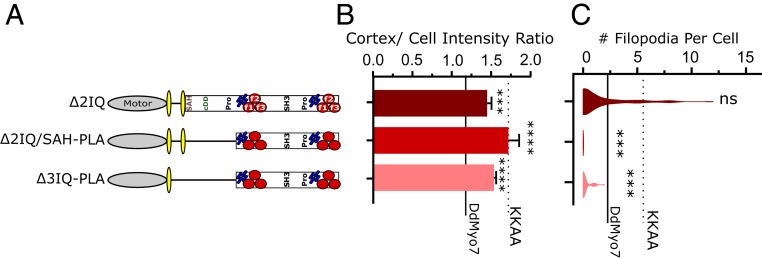
Fig. 6.
Shortening the LA and PLA disrupts filopodia formation. (A) Schematic illustration of deletion mutants. (B) Cortical intensity of LA and PLA region deletion mutants; significant values are compared to DdMyo7 control (horizontal line). (C) Violin plot of filopodia per cell. The solid horizontal line represents mean of DdMyo7 control; the dashed line represents KKAA. Significance indicators from Tukey’s test are in comparison to DdMyo7 control, unless otherwise noted: ns, not significant; ***P < 0.001; ****P < 0.0001.

