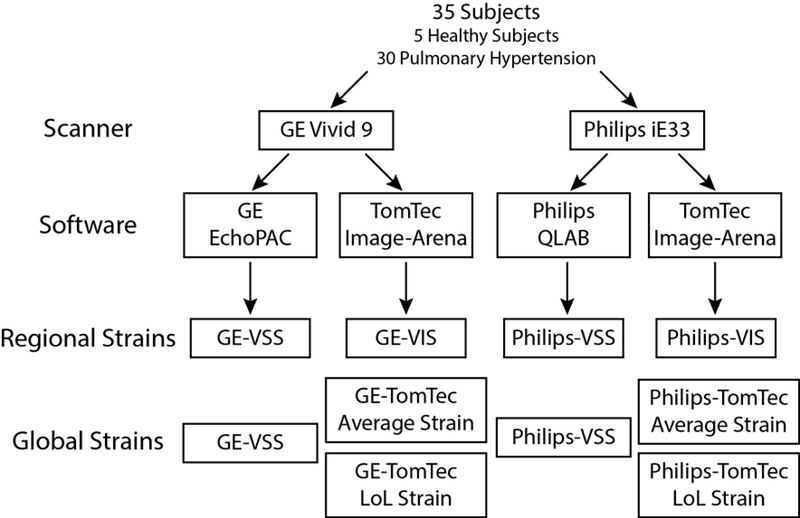
Figure 1. Study design for comparing RV regional and global strains across different scanners and software.
Thirty-five subjects had images obtained on two scanners (GE and Philips) followed by analysis in vendor-specific (VSS) and independent (VIS) software, yielding regional and global strains (see text for full details).

