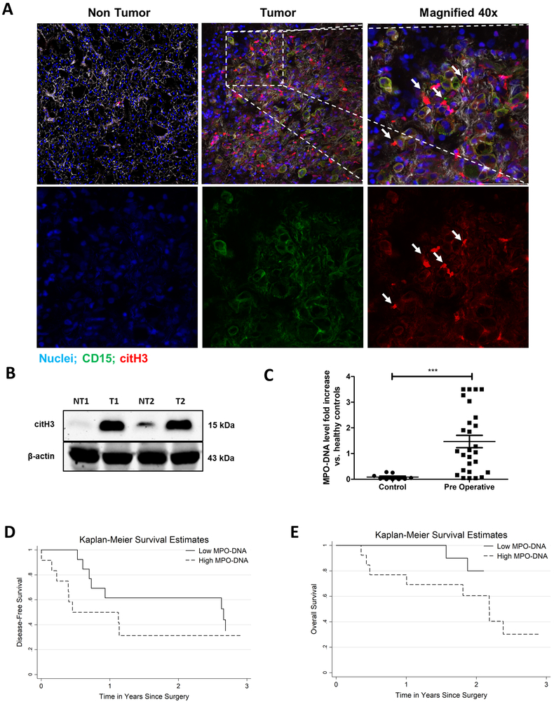
Figure 1. NET formation correlates with cancer specific outcomes in patients with metastatic colorectal cancer.
A, Representative immunofluorescence images by confocal microscopy of human colorectal liver metastases (CRLM) tissue sections showing increased neutrophil infiltration and neutrophil extracellular trap (NET) formation in tumor at 20× magnification compared to non-tumor tissue of the same patient. White arrows showing neutrophils releasing NETs in merge and single staining magnified images at 40×, scale bar 50 µm. B, Protein citH3 levels were increased as evident by western blot image between the tumor (T) and non-tumor (NT) tissue of human CRLM. The blot shown is representative of three independent experiments with similar results. C, Pre-operative MPO-DNA levels detected by Elisa kit are significantly higher in patients with CRLM (n=27) compared to healthy volunteers (n=10, ***p<0.0002). D and E, Kaplan-Meier disease-free and overall survival curves were based on high versus low MPO-DNA levels post-operatively for three years (log-rank test p<0.001 for both D and E).

