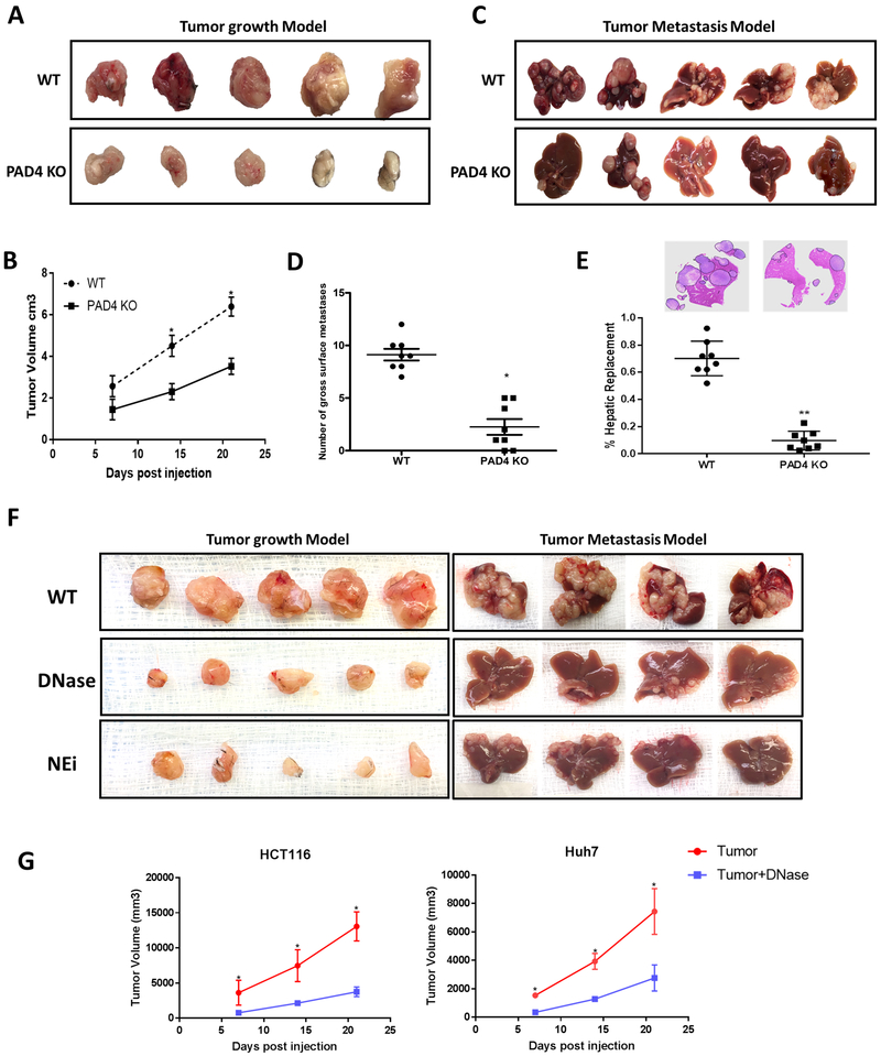
Figure 2. Tumor growth is reduced in neutrophil extracellular traps (NETs) depleted mice.
Subcutaneous tumors using MC38 cells (1×10⁶) injected subcutaneously (A) or through the spleen (C) for the metastasis model showing smaller tumors harvested 3 weeks post-inoculation in PAD4 KO compared to WT mice (n=5/group). B and D, Graphs showing significantly decreased tumor volume and surface liver nodules, respectively, in PAD4 KO mice compared to WT control *P <0.05. E, Hematoxylin and Eosin (H&E) staining of liver sections exhibit decreased tumor burden in PAD4 KO mice (n=5) compared to WT **P <0.01. F, Similarly, mice treated daily with DNAse (50ug) or Neutrophil Elastase inhibitor (NEi) (2.0 mg/kg) in both subcutaneous (n=5) and metastatic model (n=4) showed decrease tumor growth compared to controls. G, Graph representing tumor growth curve in DNAse (50ug) treated Nu/Nu athymic mice inoculated with HCT116 and Huh7 cell lines (1×10⁶) (n=5/group). Data presented as mean SEM. *P <0.05.

