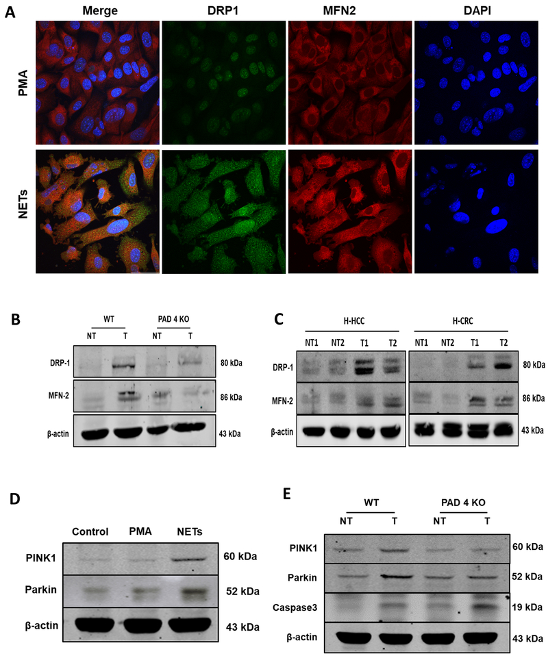
Figure 6. NETs preserve mitochondrial homeostasis and dynamics: fission, fusion and mitophagy.
A, After PMA stimulation for 4h, neutrophil extracellular traps (NETs) were cocultured with MC38 cancer cell line and confocal microscopy was utilized to analyze proteins regulating mitochondrial fission and fusion. NET treatment upregulated the expression of DRP1 and MFN2 proteins compared to cells treated with PMA as a control, nuclei (blue), DRP1 (green), and MFN2 (red). magnification 40× scale bar 50 µm. B, 3 weeks after the splenic injection the liver tumor tissue of PAD4 KO mice showing downregulated expression of DRP1 and MFN2 compared to its non-tumor background. Similarly, human hepatocellular (H-HCC) and human colorectal (H-CRC) tumor (T) sections showing upregulation in these proteins comparing non-tumor (NT) counterparts. D, Representative western blot analysis image showing increased induction of mitophagy as evident by the upregulation in the expression of protein PINK1 and Parkin in MC38 cancer cells when treated with NETs. E, Parallel results can be seen in the tumor tissues of the WT mice 3 weeks after injection as analyzed by western blot. The blots shown are representatives of three independent experiments with similar results.

