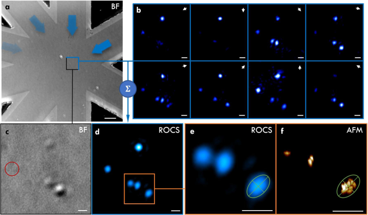
Fig. 5.
Imaging of metal-organic frameworks (MOFs) with ROCS. (a) Overview of the imaged region. (b) Raw evanescent scattering images under waveguide illumination with a 488 nm laser. (c) Brightfield image using sum of multiple LED wavelengths. The red circle highlights a cluster that is only visible under darkfield illumination. (d) Intensity image created by ROCS with (e) zoom onto MOF clusters. Although it is not possible to discern individual particles, the elongated shape of the clusters is visualised by ROCS in good agreement with (f) the atomic force microscopy image of the same region. The overview image (a) measures 100 × 100 μm2 and the scalebars in (b–f) are 1 μm.

