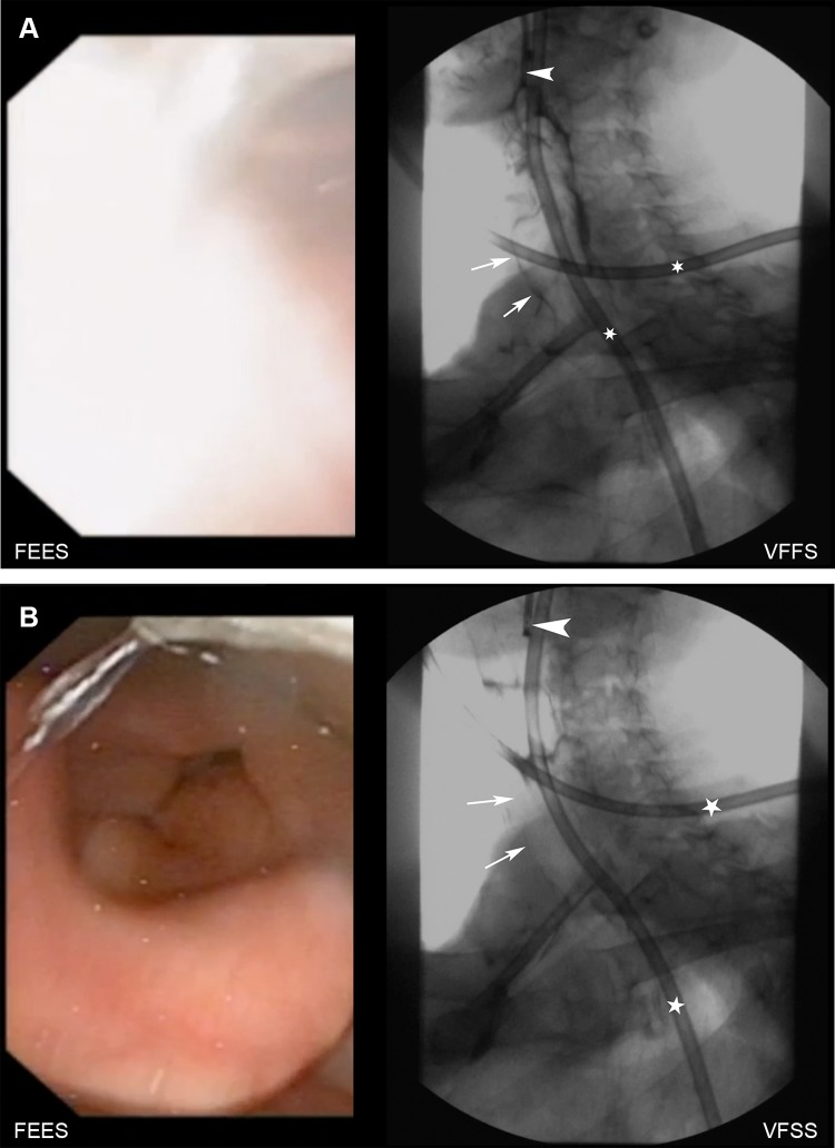
Fig. 2.
a shows the swallow of a 5 ml bolus of liquid consistency. On the left, an intradeglutitive “white-out” is seen on FEES, VFSS shows intradeglutitive silent aspiration (arrows; arrowhead: endoscope, asterisks: nasogastric tube). b shows same patient after swallowing: no intralaryngeal or intratracheal contrast medium is seen on FEES as well as on VFSS (arrows, arrowhead: endoscope, asterisks: nasogastric tube)

