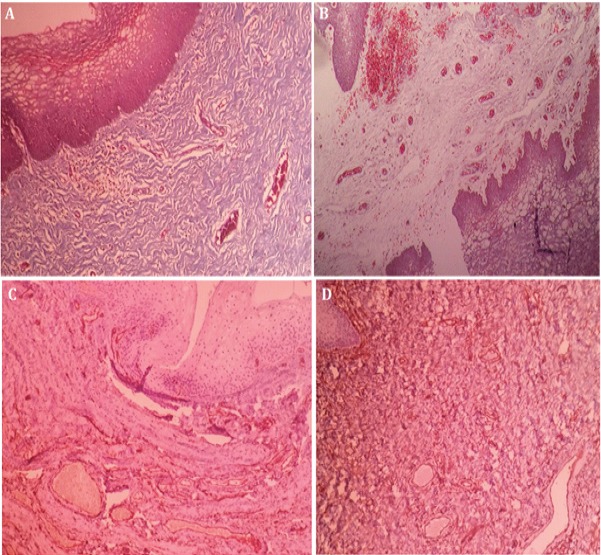
Figure 2.

Lateral Wall Vaginal Biopsy Stained with Masson’s Trichrome Staining and IHC CD34) for Evaluation of Angiogenesis in 35 Years Women with Rectum Cancer that She has not Able to Intercourse Over the Last Three Years (after irradiation) due to Severe Vaginal Atrophy and Stenosis. A and C are belonged to this patient before receiving APRGF and B and D are belonged to the same patient after receiving three doses of the ARPGF. A: showed subepithelial dense fibrosis (40×). B: Subepithelial loose fibroconnective tissue without fibrosis (40×). C and D: there is no significant difference before and after APRGF injection in IHC staining for CD34 in this biopsy section (40×).
