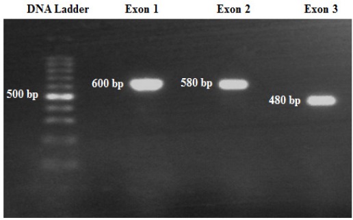
Figure 1.

Representative Gel Electrophoresis of NUDT15 Gene’s Exons after PCR. Ten µl of PCR product was run on 1.5% agarose gel and stained with Redsafe dye. The bands were visualized by exposing the gel to UV light using the bench top U.V transluminator (Bio Doc-ITTM, UK). Further information are presented in the Materials and Methods section.
