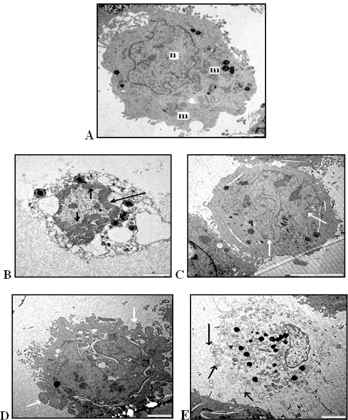
Figure 7.

Transmission Electron Micrographs of DBTRG.05MG Cells at Various Stages of Apoptosis (A) untreated DBTRG.5MG cell with intact nucleus (n) mitochondria (m) and clear cytoplasm (Magnification 8000X).(B) chromatin condensed at the nuclear periphery (arrow) (Magnification 8000X). (C) Apoptotic cell containing nuclear fragments (arrow) (Magnification 8000X). (D) Membrane blebbing indicated by arrow (Magnification 12000X).
