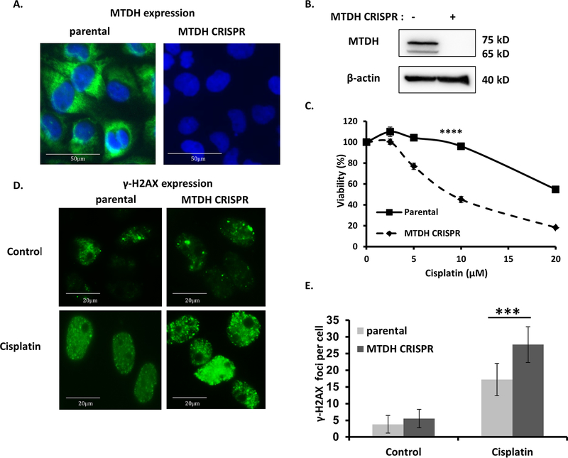
Fig. 4. MTDH depletion increases sensitivity to cisplatin and accumulation of DNA damage in cancer cells.
(A) Fluorescent imaging of MTDH (green) and nuclei (blue, DAPI) in control and MTDH CRISPR knockout cancer cells. (B) Western blot confirming deletion of MTDH using CRISPR/Cas9 in Hec50 cells. β-tubulin is the loading control. (C) Sensitivity to cisplatin was examined in parental and MTDH CRISPR knockout Hec50 cells by the WST-1 assay from three independent experiments. ****P<0.0001 by two-way ANOVA. (D) Immunostaining was used to detect γ-H2AX foci in parental and MTDH CRISPR knockout Hec50 cells without treatment or treatment with 5 µM cisplatin for 16 h. (E) Quantification of γ-H2AX foci in cancer cells. Data are representative of 300 cells from three independent experiments, ***P<0.001.

