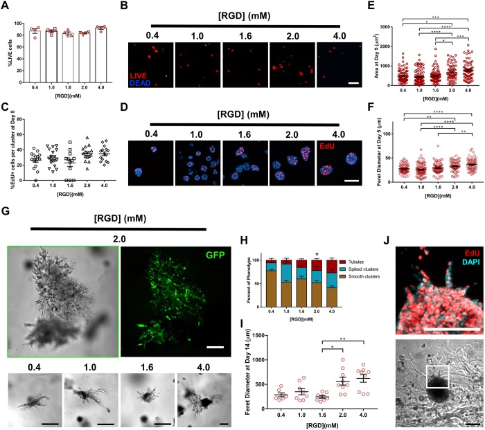
Fig. 6.
Adhesive peptide density in PEG-4MAL hydrogels regulates tubule formation. (A) Percentage of IMCD cells that stained for LIVE (C12-Resazurin) after 1 day of encapsulation in 8%, 20 kDa PEG-4MAL hydrogels functionalized with varying RGD density and crosslinked with IPES peptide (mean±s.e.m.). Each data point represents one independent hydrogel. At least 100 cells were assessed per condition. (B) Fluorescence microscopy images of IMCD cells cultured in PEG-4MAL hydrogel. IMCD cell viability was assessed at 1 day after encapsulation. Scale bar: 50 μm. (C) Percentage of IMCD cells per cluster that were labeled by EdU incorporation (mean±s.e.m.) after 5 days of encapsulation. At least 30 clusters were analyzed per condition. (D) Fluorescence microscopy images of proliferating (EdU+) IMCD cells cultured in PEG-4MAL hydrogels of different polymer density. IMCD cell proliferation was assessed at 5 days after encapsulation. Scale bar: 50 μm. IMCD multicellular structure (E) projected area and (F) Feret diameter at 5 days after encapsulation in PEG-4MAL hydrogel. Graph line represents the mean of the individual data points. Each data point represents one multicellular structure. (G) Transmitted light and fluorescence microscopy images of GFP-expressing IMCD cells at 14 days after encapsulation in PEG-4MAL hydrogel. Scale bars: 100 μm. (H) Percentage of IMCD multicellular structures (mean±s.e.m.) that were classified as ‘smooth clusters’, ‘spiked clusters’ or ‘tubules’ after 14 days of encapsulation. At least 10 multicellular structures were analyzed per condition. *P<0.0021 for 2.0 versus 0.4 mM RGD (χ2 test with Bonferroni's correction). (I) IMCD multicellular structure Feret diameter at 14 days after encapsulation in PEG-4MAL hydrogel. Graph line represents the mean of the individual data points. Each data point represents one multicellular structure. ****P<0.0001, ***P<0.0002, **P<0.0021, *P<0.0332 (Kruskal–Wallis with Dunn's multiple comparisons test). (J) Transmitted light and fluorescence microscopy images (magnification of boxed area) of proliferating (EdU+) IMCD cells cultured in PEG-4MAL hydrogels functionalized with 2.0 mM RGD. Scale bars: 100 μm. Experiments performed with six PEG-4MAL hydrogels per experimental group. Three independent experiments were performed and data are presented for one of the experiments.

