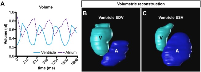
Fig. 5.
Volumetric reconstructions using CFIN. (A) Continuous chamber volumes over three cardiac cycles in a wild-type 48 hpf zebrafish embryo. Volumes were calculated using synchronized images processed by CFIN. Ventricle (cyan); atrium (purple dashed). (B,C) Graphical 3D reconstruction of ventricular EDV (B) and ESV (C) as measured by CFIN. Ventricle (V); atrium (A).

