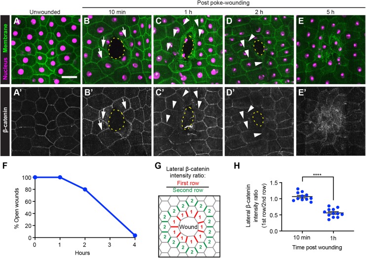
Fig. 1.
Junctional β-catenin in wound-edge epidermal cells is reduced after wounding. (A-E′) Dissected epidermal whole mounts of unwounded (A,A′) or poke-wounded (B-E′) third instar larvae expressing UAS-DsRed2nuc (nuclei, magenta) and UAS-src-GFP (cell membranes, green) via the A58-Gal4 driver 10 min (B,B′), 1 h (C,C′), 2 h (D,D′) and 5 h (E,E′) after wounding. (A-E) The nuclei and cell membrane. (A′-E′) The adherens junctions of the same samples immunostained using anti-β-catenin antibodies (white). Scale bar: 50 μm. Dotted yellow lines indicate wound borders. Arrows in B,B′ highlight examples of clear junctional β-catenin signal (B′) and membrane-GFP signal (B). Arrowheads in C-D′ highlight examples of reduced junctional β-catenin (C′,D′) where membrane-GFP is still present (C,D). (F) Quantitation of open poke wounds in control larvae. The epidermal reporter used was e22c-Gal4, UAS-LifeAct-Cherry, UAS-luciferaseRNAi. n≥20 for each time point. (G) Schematic of quantitation strategy for measuring β-catenin levels on lateral segments near the wound. (H) Quantitation of junctional β-catenin ratio in first- and second-row wound-edge epidermal cells. Each dot represents one larva. Data are mean±s.e.m. ****P<0.0001 (unpaired t-test).

