Fig. 1.
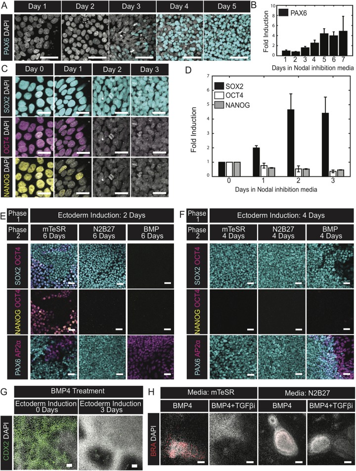
During neural differentiation cells transition through an ectodermal progenitor state before commitment. (A,C) Representative images of cells fixed on the indicated days during the course of Nodal inhibition and immunostained for the neural progenitor marker PAX6 (A) or the pluripotent markers SOX2, OCT4 and NANOG (C). (B,D) Quantification of the fold change in average intensities of PAX6 (B) or SOX2, OCT4 and NANOG (D) normalized to DAPI for the indicated days relative to day 0. N=3 experiments. (E,F) Representative images of cells immunofluorescently co-stained for AP2α/PAX6 or SOX2/NANOG/OCT4 at the conclusion of a two-phase induction protocol. Cells were initially differentiated in Nodal inhibition media for either 2 (E) or 4 (F) days and then treated with the indicated culture conditions for the subsequent 6 (E) or 4 (F) days. Experiment replicated three times. (G) Representative images of cells immunofluorescently labeled for CDX2. All cells were treated with BMP4 for 2 days following either no prior treatment or 3 days of Nodal inhibition. Experiment replicated three times. (H) Representative images of cells immunofluorescently labeled for BRA at the conclusion of the experiment. All cells were initially induced for 3 days in Nodal inhibition media. Thereafter, the media was exchanged for treatment conditions indicated in the banner above the corresponding images. First row banners indicate the base media and the second banner row indicates the signals added to the base media. Experiment replicated three times. TGFβi, SB431542. Data are mean±s.e.m. Scale bars: 25 µm (C); 50 µm (A,E,F); 100 µm (G,H).

