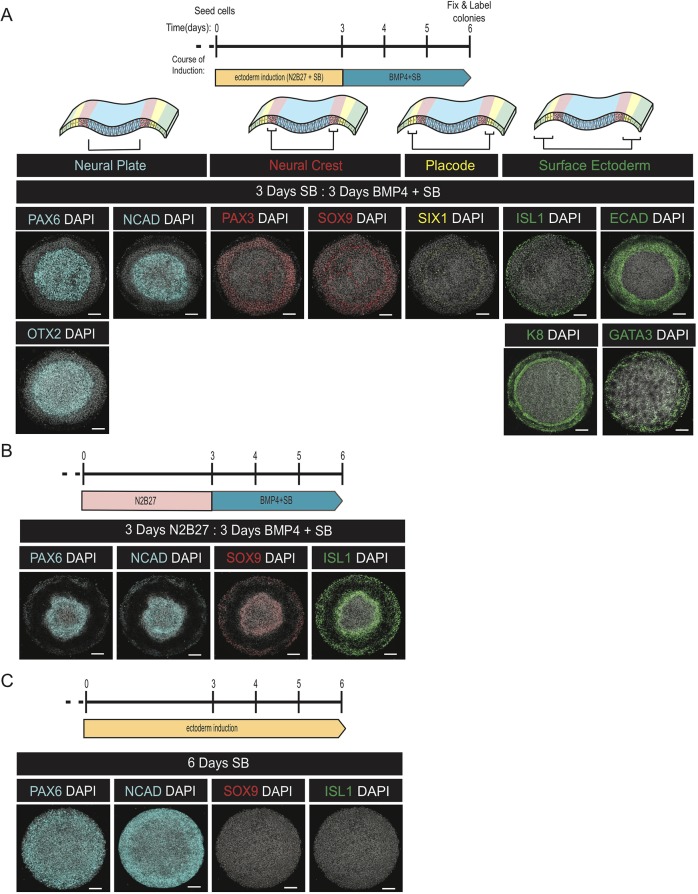
Fig. 2.
A two-phase protocol generates self-organized patterns of ectodermal fates. (A-C) Representative images of colonies stained with the indicated antibodies following treatment with either a two-step protocol consisting of 3 days in ectoderm induction media and 3 days in N2B27+BMP+SB (A), the same protocol except the first 3 days were in N2B27 alone (B), or 6 days of ectoderm induction media (C). The top row in (A) shows a schematic of the organization of fates in the anterior ectoderm. Each experiment was replicated at least three times. Scale bars: 100 μm.

