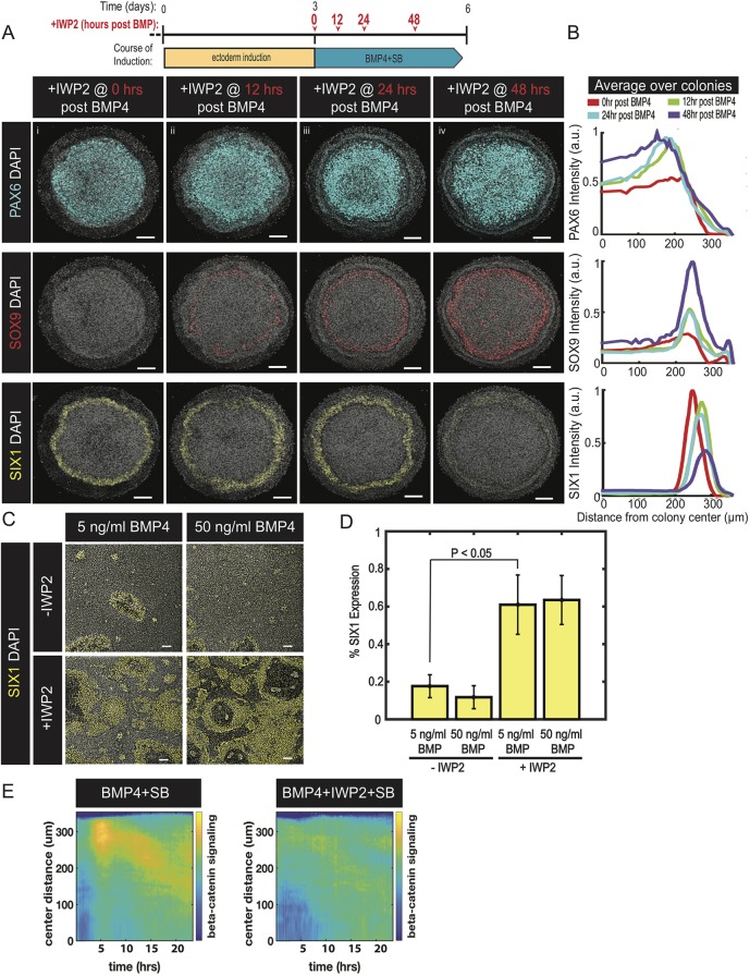
Fig. 3.
Endogenous WNT ligands drive differentiation to neural crest at the expense of placodal fates. (A) Representative images of colonies immunostained for PAX6, SOX9 or SIX1. Colonies were initially induced for 3 days in ectoderm induction media and then subsequently differentiated for 3 days in N2B27 media containing BMP4 and SB. The time between BMP4 and IWP2 addition is indicated in the induction schematic and linked in red with the banner above the corresponding image. Experiment replicated four times. (B) Quantification of the images in A represented as average nuclear intensities of the indicated markers normalized to DAPI as a function of radial position. N=3 colonies. (C) Representative images of cells in standard culture immunostained for SIX1. Cells were initially induced for 3 days in ectoderm induction media and then treated with either 5 or 50 ng/ml of BMP4 in media with (+) or without (−) IWP2 for the subsequent 3 days. (D) Fraction of cells expressing SIX1. N=3 experiments; P=0.011 (two-sided t-test). Data are mean±s.e.m. (E) Kymograph of β-catenin signaling activity over a 24-h period (on day 4) in micropatterned colonies initially treated in Nodal inhibition media for 3 days and then with the indicated signaling conditions. N=4 colonies and repeated twice. Colony diameter in A: 700 µm. Scale bars: 100 µm.

